
Science
We are pioneering the future of non-invasive neuroimaging. By moving beyond traditional Functional Near-Infrared Spectroscopy (fNIRS), we have delivered high-resolution, functional brain imaging: High-Density Diffuse Optical Tomography (HD-DOT).
Explore the fundamental science behind our technology below.
Functional near-infrared spectroscopy (fNIRS)
When neurons are active, their metabolic demands increase. Local blood vessels respond by dilating, leading to an influx of blood to the surrounding tissue. The relationship between neuronal activity and the localised response of the blood vessels is known as neurovascular coupling.
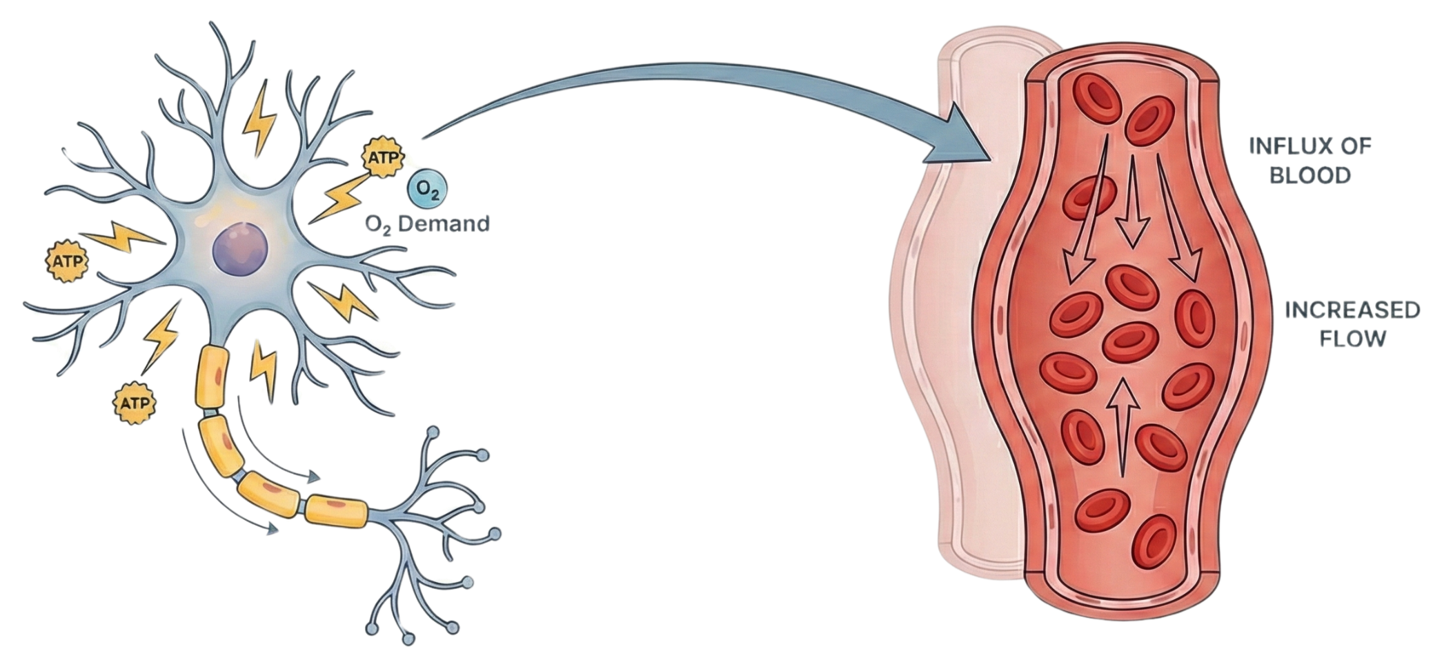
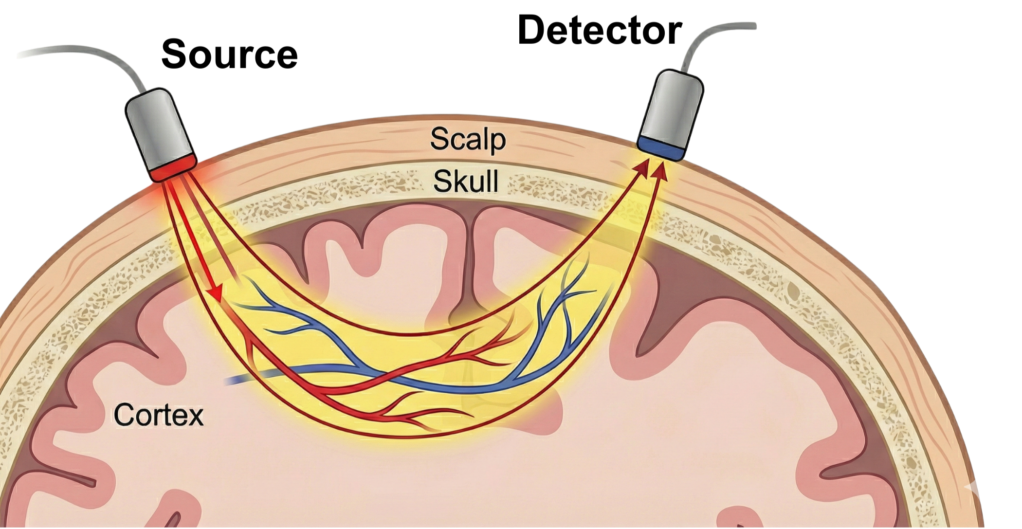
Therefore, by transmitting near-infrared light at two wavelengths into the scalp and detecting the light that scatters back to the surface, it is possible to measure changes in haemoglobin concentration in the cortex and, from this, infer brain activity.
This non-invasive neuroimaging technique is called functional near-infrared spectroscopy(fNIRS).
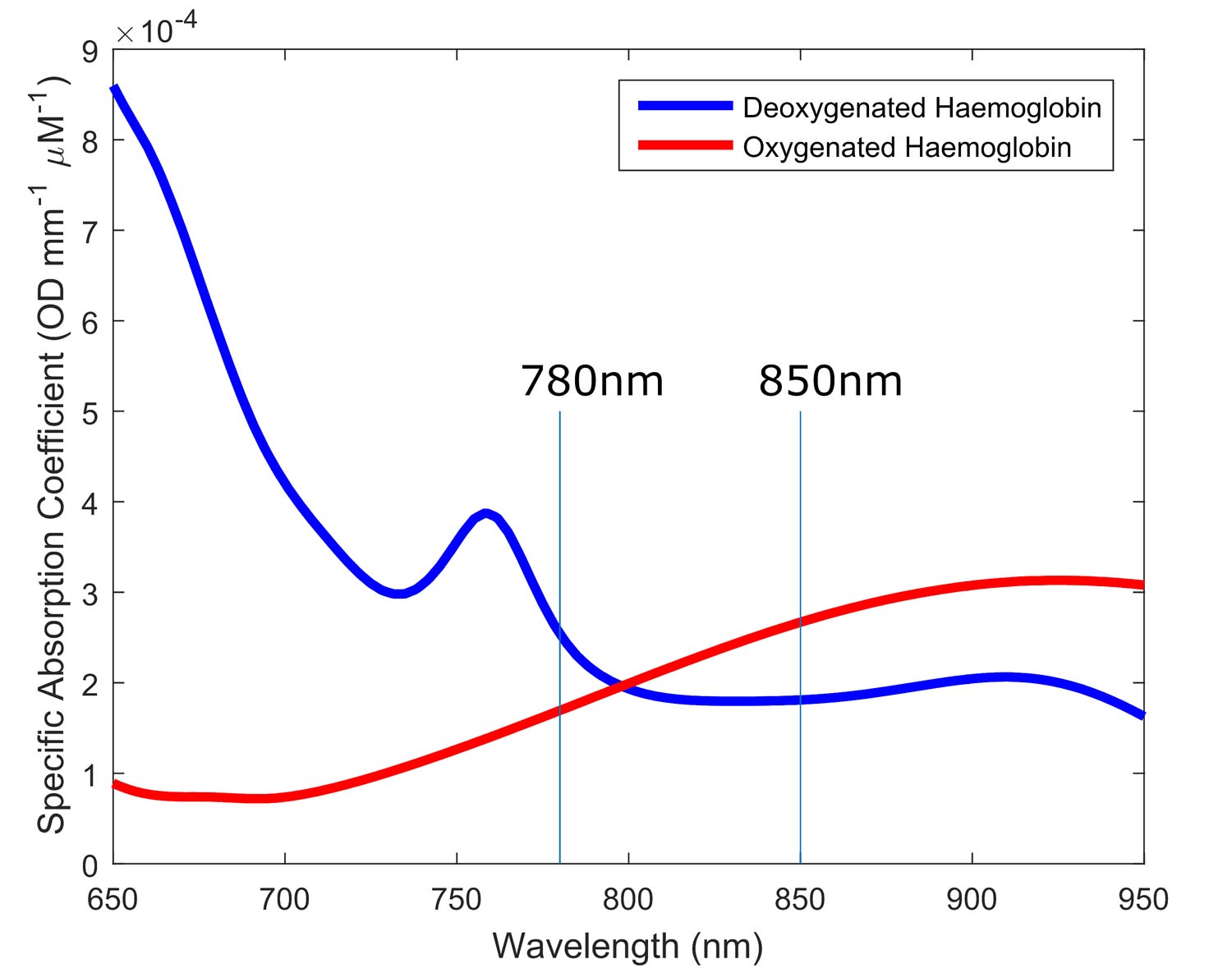
Human tissue is relatively transparent to red and near-infrared light, while haemoglobin absorbs light at these wavelengths - and critically, oxygenated and deoxygenated haemoglobin have different absorption spectra.
High-density diffuse optical tomography (HD-DOT)
A basic fNIRS measurement consists of a source-detector pair, known as a channel. Each channel is sensitive to changes in blood oxygenation up to a maximum depth of around one-third of the separation between its source and detector.
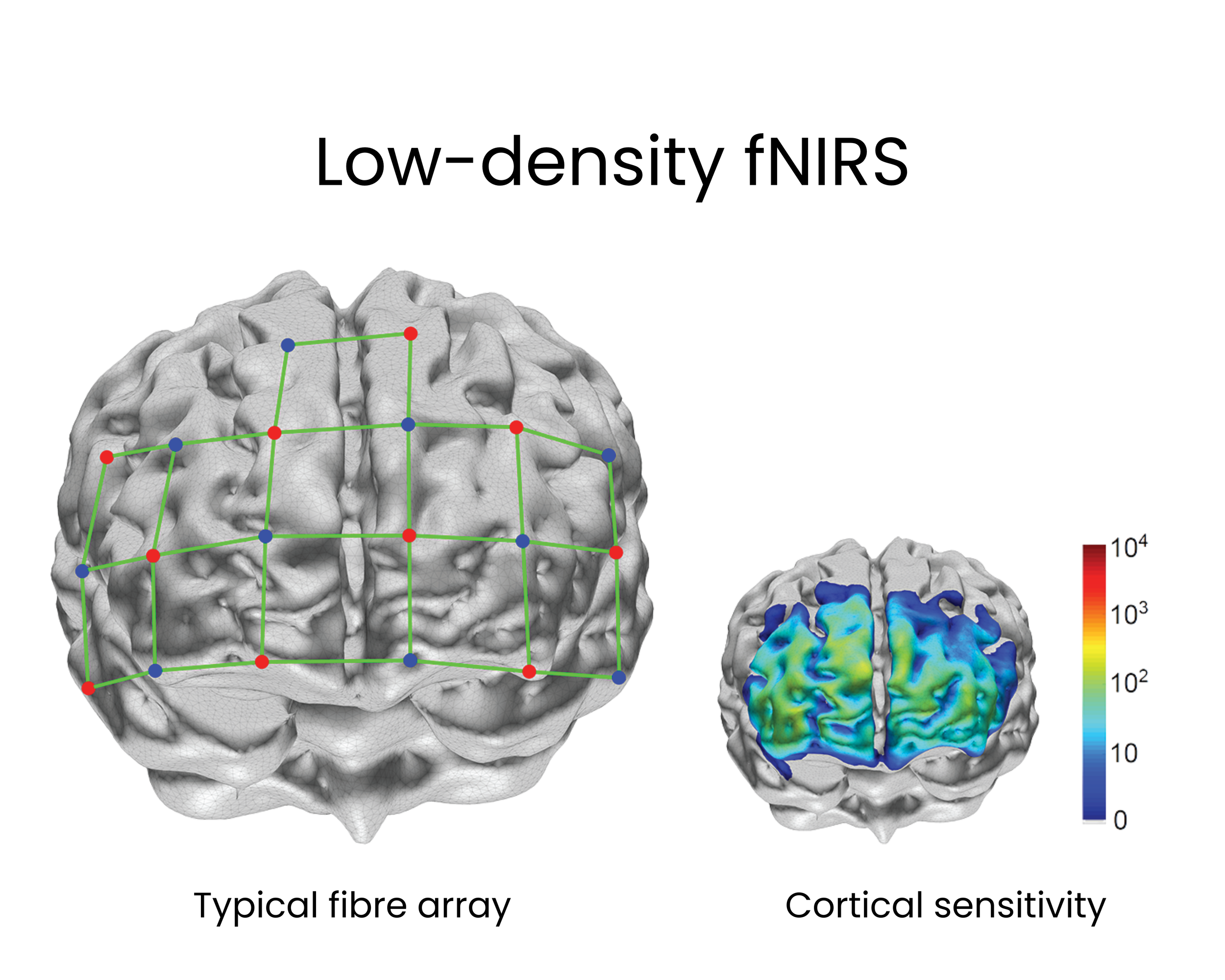
Low-density fNIRS systems are limited to 10 to 60 non-overlapping channels, using a fixed source-detector distance (typically 30 mm for adults). As a result, spatial resolution is limited, forcing researchers to rely on basic channel-by-channel data analysis.
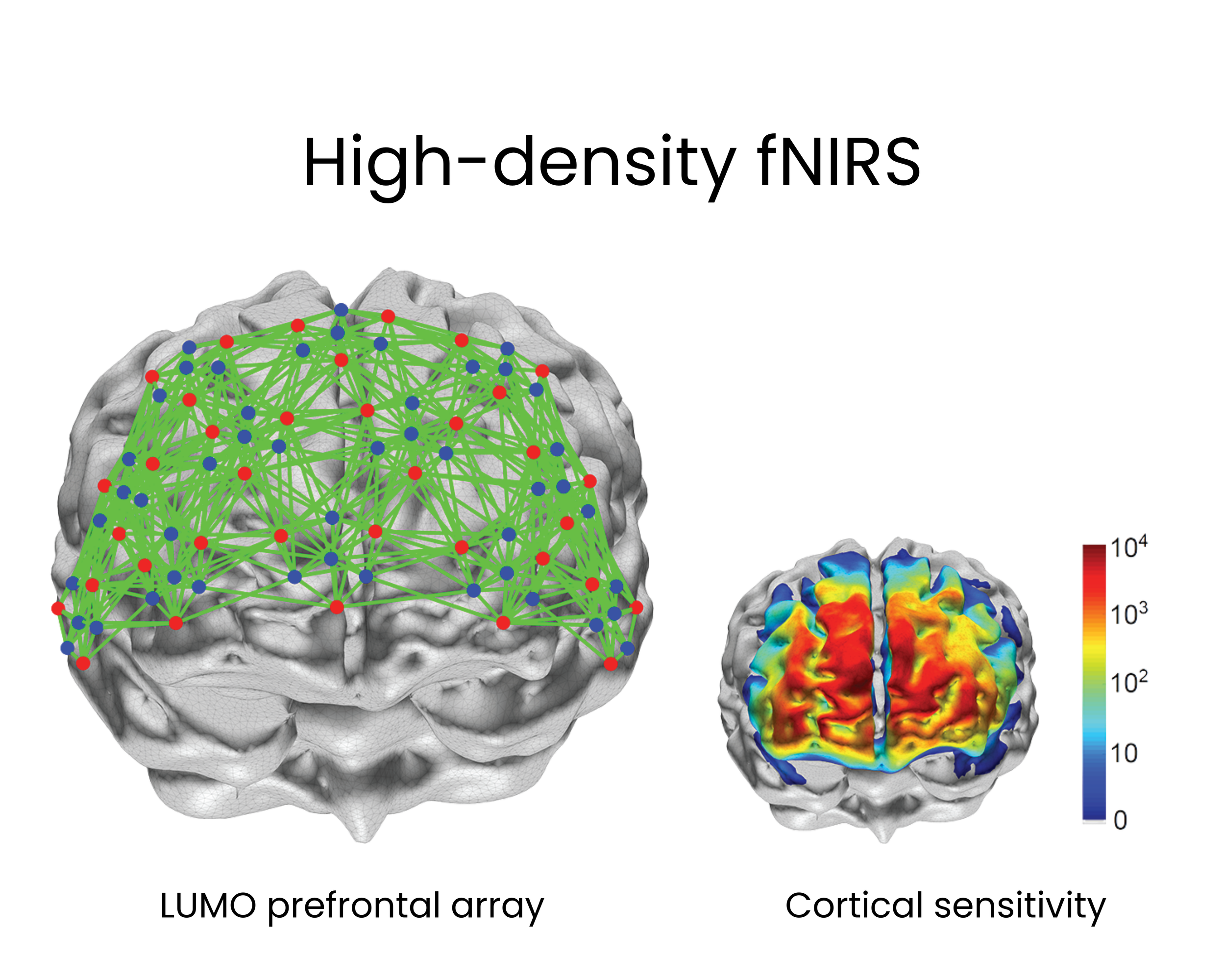
A standard LUMO12 prefrontal array (right) provides over 1,200 dual-wavelength channels, with overlapping source-detector separations ranging from 10 mm to 70 mm.
A whole-head LUMO54 system yields approximately 7,000 channels.
LUMO’s dense, overlapping network of thousands of fNIRS measurements at multiple separations contains rich spatial information.
Diffuse optical tomography (DOT) is an imaging modality that fully exploits this data.
Using a forward model of light propagation and an inverse reconstruction algorithm, high-density DOT (HD-DOT) transcends traditional channel-by-channel analysis - reconstructing precise, three-dimensional images of functional activation in the cortex.

Further details on DOT theory and applications can be found in these selected articles:
Functional brain mapping using whole-head very high-density diffuse optical tomography.
Fogarty et al (2025)Diffuse optical tomography in the human brain: A brief review from the neurophysiology to its applications.
Hernández-Martín & González-Mora (2020)High-density diffuse optical tomography for imaging human brain function.
Wheelock et al (2019)Mapping distributed brain function and networks with diffuse optical tomography.
Eggebrecht et al (2014)Optical tomography: Forward and inverse problems.
Arridge & Schotland (2009)